
Translate this page into:
Multiple diastema closure with lithium disilicate veneers

*Corresponding author: Ashok Rupa, Department of Conservative Dentistry and Endodontics, Sri Ramachandra Institute of Higher Education and Research, Chennai, Tamil Nadu, India. rupa@sriramachandra.edu.in
-
Received: ,
Accepted: ,
How to cite this article: Hari R, Rupa A, Karthick S, Mathan Rajan R. Multiple diastema closure with lithium disilicate veneers. Sri Ramachandra J Health Sci. 2024;4:68-71. doi: 10.25259/SRJHS_35_2023
Abstract
Multiple diastema is one of the most prevalent esthetic issues that significantly reduces the patient’s confidence. This condition still poses a challenge for a clinician to restore these gaps and to provide an esthetic smile which is desired by the patient. Indirect Veneer is one of the treatment options for the closure of diastema which provides a predictable result. This case report describes the management of multiple diastema in maxillary anterior teeth with lithium disilicate veneer. A 40-year-old female patient reported to the department of Conservative Dentistry and Endodontics of Sri Ramachandra Dental College, Sri Ramachandra Institute of Higher Education and Research with a chief complaint of gaps in her upper front teeth. All treatment options were evaluated and discussed with the patient and the patient agreed on an indirect veneer for diastema closure. The treatment involved a complete digital workflow with direct intra-oral scanning, digital smile designing, and computer-aided milling. Indirect veneer for multiple diastema closure using a complete digital workflow provides predictable and satisfactory results.
Keywords
Digital smile designing
Multiple diastema
Veneer

INTRODUCTION
Multiple diastema is a type of malocclusion that can hinder a person’s self-confidence regarding their smile and appearance. Its etiology varies from dental malformations, macroglossia, genetics, and habits.[1] Veneers were introduced as a potential restorative modality. The acid etch technique and silica resin direct filling material led to an increase in interest toward laminate veneers.[2] Veneers as an esthetic solution for diastema pose a challenge in clinical practice being bonded only to the facial surface of anterior teeth using resin cements.[3] This procedure is highly conservative and produces excellent esthetic results. They take up the strength of enamel when they are bonded to it and become as strong as the teeth. This case report describes the closure of multiple diastema in maxillary anterior teeth with lithium disilicate (LiDi) veneers using an entirely digital workflow.
CASE REPORT
A 40-year-old female patient reported to the department of Conservative Dentistry and Endodontics of Sri Ramachandra Dental College, Sri Ramachandra Institute of Higher Education and Research with a chief complaint of gaps in her upper front teeth. On examination, diastema was found between 11, 12, 13, 21 22, and 23 with Ellis and Davey’s Class I fracture in 11 [Figure 1a-d]. On cold test, normal response was elicited in all the maxillary anterior teeth. The patient was explained all the treatment options and intra-oral and extra-oral photographs were taken on the first visit and pre-operative shade was evaluated [Figure 1e]. A complete digital workflow for the fabrication of the veneers was opted.
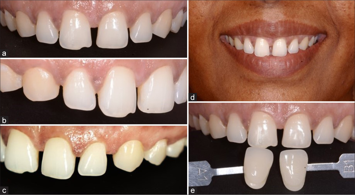
- Pre-operative (a) frontal view, (b) right lateral view, (c) left lateral view, (d) smile line evaluation, and (e) shade evaluation.
On the second visit, a pre-operative occlusal evaluation was done [Figure 2a] and then a direct intra-oral digital impression was taken using the i700 (Medit) scanner [Figure 2b]. The maxillary teeth, mandibular teeth, and occlusion were recorded [Figure 2c]. Using the recorded data, digital smile designing was done [Figure 2d] and a mock-up was 3D printed [Figure 2e].
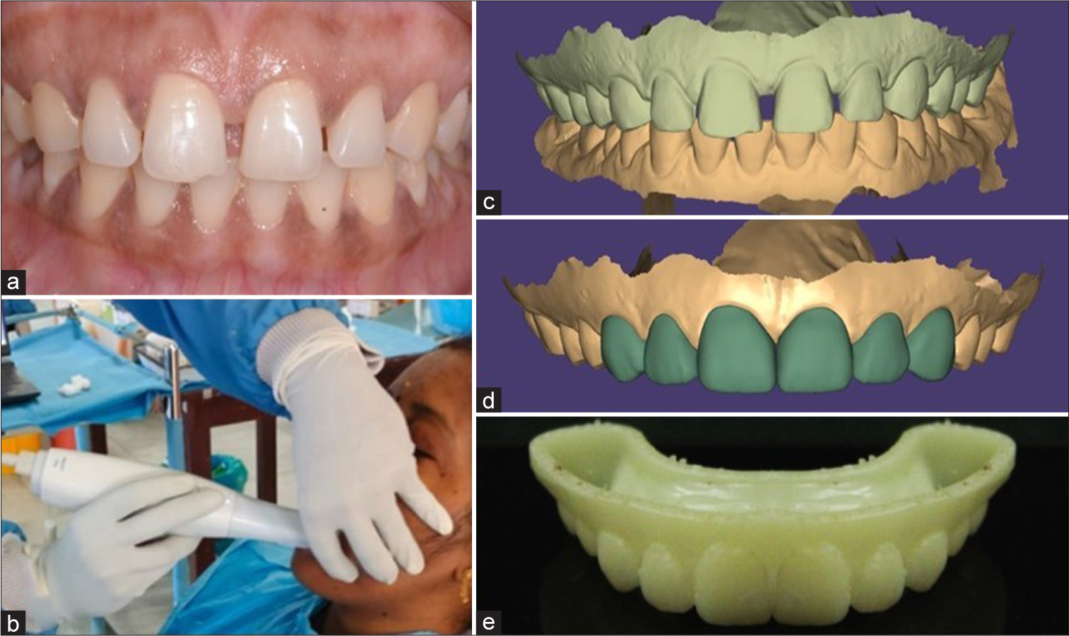
- (a) Pre-operative occlusion evaluation, (b) intra-oral scanning, (c) scanned data, (d) digital smile designing, and (e) 3D printed mock-up.
On the third visit, the mock-up was transferred to the mouth using Structur 2 (VOCO). Once the patient is satisfied with the mock-up, veneer preparation was done from canine to canine to a depth of 0.5 mm with incisal butt joint preparation and equigingival margin using Mani Depth Marker Dia bur DM-305. A direct digital impression was taken and sent to the lab for veneer fabrication [Figure 3a]. The teeth were temporized using bis-acryl composite using the spot-etch technique [Figure 3b-d].
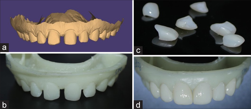
- (a) Digital scan of tooth preparation for veneer, (b) 3D printed cast, (c) fabricated veneers, and (d) veneers in 3D printed cast evaluating the fit.
On the fourth visit, the temporary veneers were removed, and a try-in paste was used to try the LiDi veneer and checked for marginal adaptation, color, contact, and occlusion. Surface treatment was carried out on veneers of LiDi by application of 9% hydrofluoric acid for 20 s, rinsed, and dried [Figure 4a and b]. This was followed by acid etching with 37% phosphoric acid for 60 s, rinsed, and dried [Figure 4c]. Silane was applied to the intaglio surface of the veneer for 60 s [Figure 4d-f]. Simultaneously, the tooth surface was etched with 37% phosphoric acid for 30 s, rinsed, and dried [Figure 4g and h]. A bonding agent was applied with a micro brush [Figure 4i]. LiDi veneer cementation was done using resin cement (G-Cem linkforce, GC) [Figure 5a-d]. Post-operative instructions were given.
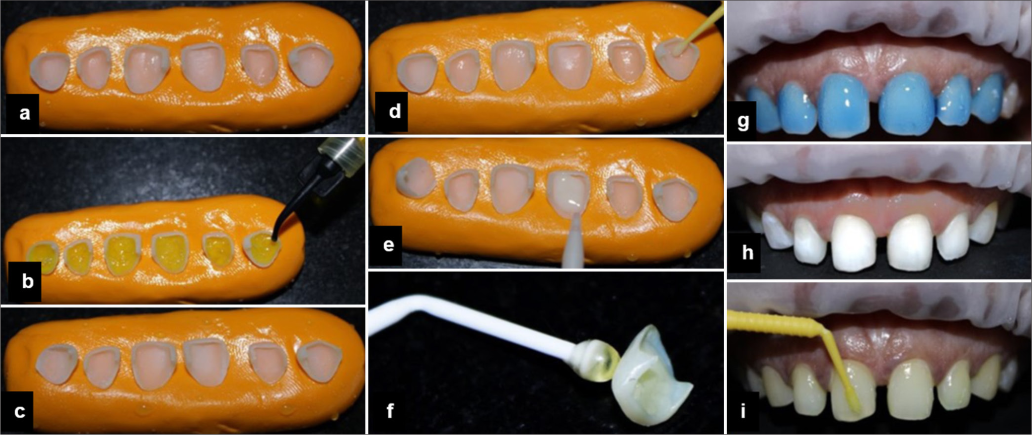
- (a) Veneers stabilized in elastomeric impression material, (b) veneers etched with 9% hydrofluoric acid, (c) etched veneer surface, (d) silanizing the veneers, (e) resin applied onto the veneer, (f) veneers carried using sticky applicator tip, (g) teeth etched with 37% phosphoric acid, (h) etched teeth, and (i) bonding agent applied onto the teeth.
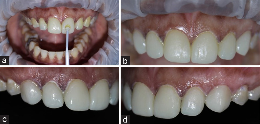
- (a) Veneers transferred to the teeth using sticky applicator tip, post-operative (b) frontal view, (c) right lateral view, and (d) left lateral view.
During the 1-week follow-up, the patient gave positive feedback, there were no extra-oral abnormalities, the intra-oral examination revealed good color stability of the veneers, and the gingival health was optimal [Figure 6a-d].
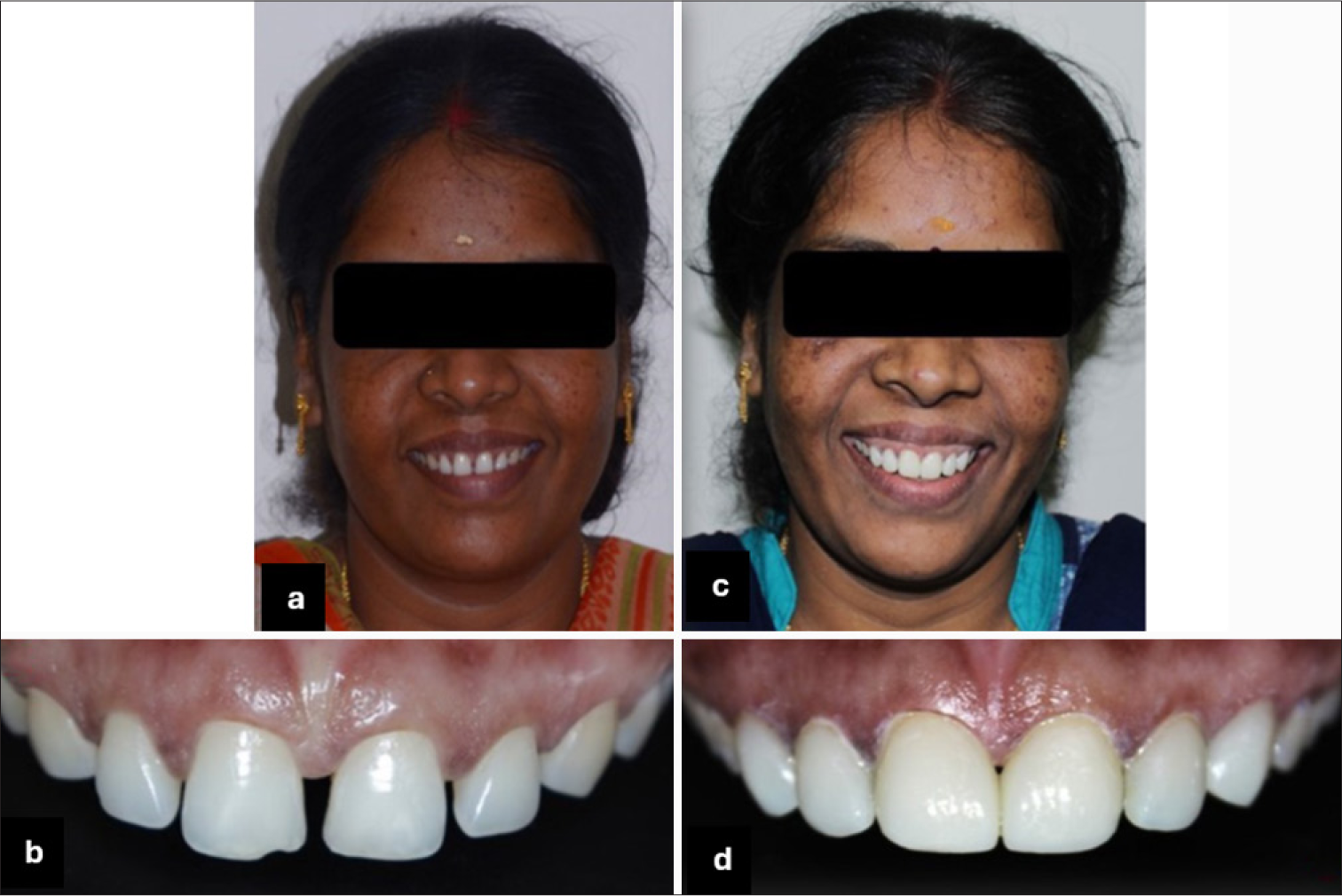
- Pre-operative (a) face, (b) teeth, post-operative (c) face , and (d) teeth.
DISCUSSION
Diastema is an esthetic problem that occurs more frequently in the maxilla than the mandible[4] with a higher prevalence in women compared to men and more common in the age range of 12–19 years.[5] In this case, during treatment planning, a digital smile design (DSD) and mock-up was made. DSD is a practical tool which helps in esthetic diagnosis, better communication between patient, clinician and laboratory, predicting each phase of treatment, increase patient knowledge, and motivation.[6] In spite of its many advantages, mock-up is still needed as an efficient way for communicating treatment plans and also serving as a tool to confirm results from digital workflow.[7] Treatment of diastema can be done by orthodontic approach, restorative approach, surgical approach, or a multidisciplinary approach based on the cause.[8]
LiDi veneer restoration is a popular treatment modality for closing diastema due to its highly esthetic properties, exceptional color stability, and better mechanical properties than composite resins.[9] LiDi is a glass ceramic with a 70% ceramic crystal concentration.[10] Its unique microstructure is key to their mechanical and optical properties. The survival rate of LiDi was reported to be 98.7% at the end of 3 years. and 97.4% at the end of 10 years, among those 85.5% of the restorations were plaque-free, with no significant color change and surface characteristics of the veneers.[11] LiDi was the choice of material not only due to its superior mechanical properties but also due to its exceptional optical properties, making it as the most appropriate choice for the anterior region, thereby enabling the clinician in achieving a highly esthetic without compromising on the function.
CONCLUSION
Determining the etiology, analyzing the clinical features and establishing an appropriate treatment plan can determine the success and prognosis of treatment. The use of a complete digital workflow helps in the avoidance of cumulative error which occurs during the conventional workflow. The choice of LiDi in the anterior region provides a more esthetically pleasing result.
Ethical approval
The Institutional Review Board approval is not required.
Declaration of patient consent
The authors certify that they have obtained all appropriate patient consent.
Conflicts of interest
There are no conflicts of interest.
Use of artificial intelligence (AI)-assisted technology for manuscript preparation
The authors confirm that there was no use of artificial intelligence (AI)-assisted technology for assisting in the writing or editing of the manuscript and no images were manipulated using AI.
Financial support and sponsorship
Nil.
References
- Etiological factor of midline diastema-a retrospective study. Int J Dent Oral Sci. 2021;8:4119-23.
- [CrossRef] [Google Scholar]
- Conservative and esthetic management of diastema closure using porcelain laminate veneers. Med J Armed Forces India. 2015;71:S581-5.
- [CrossRef] [PubMed] [Google Scholar]
- Sturdevant's art and science of operative dentistry. Vol 5. United States: Mosby; 2006. p. :807-40.
- [Google Scholar]
- Minimum intervention management of diastema closure using cordless displacement system and laminate veneers: A 2-year follow-up. Eur J Dent. 2018;12:446-9.
- [CrossRef] [PubMed] [Google Scholar]
- Retrospective evaluation of the prevalence of diastema among an adult population. Open J Stomatol. 2022;12:175-82.
- [CrossRef] [Google Scholar]
- Digital smile design: A digital tool for esthetic evaluation, team communication, and patient management In: Ronald E, ed. Goldstein's esthetic in dentistry (3rd ed). United States: John Willey and Sons. Inc.; 2018. p. :85-111.
- [CrossRef] [Google Scholar]
- Digital smile design and mock-up technique for esthetic treatment planning with porcelain laminate veneers. J Conserv Dent. 2018;21:455-8.
- [CrossRef] [PubMed] [Google Scholar]
- Closure of midline diastema by composite resin build-up: A case report. Indian J Public Health Res Dev. 2019;10:2845-7.
- [CrossRef] [Google Scholar]
- Color stability and translucency of ceramic laminate veneer restoration for diastema closure: Effect of resin cement containing different photoinitiator. Research Square; 2022:1-24.
- [CrossRef] [Google Scholar]
- Clinical and esthetical evaluation of 79 lithium disilicate multilayered anterior veneers with a medium follow-up of 3 years. Eur J Dent. 2019;13:581-8.
- [CrossRef] [PubMed] [Google Scholar]
- Retrospective analysis of lithium dissilicate laminate veneer applied by experience dentist: 10-years result. Int J Prostodont. 2019;32:471-4.
- [CrossRef] [PubMed] [Google Scholar]


